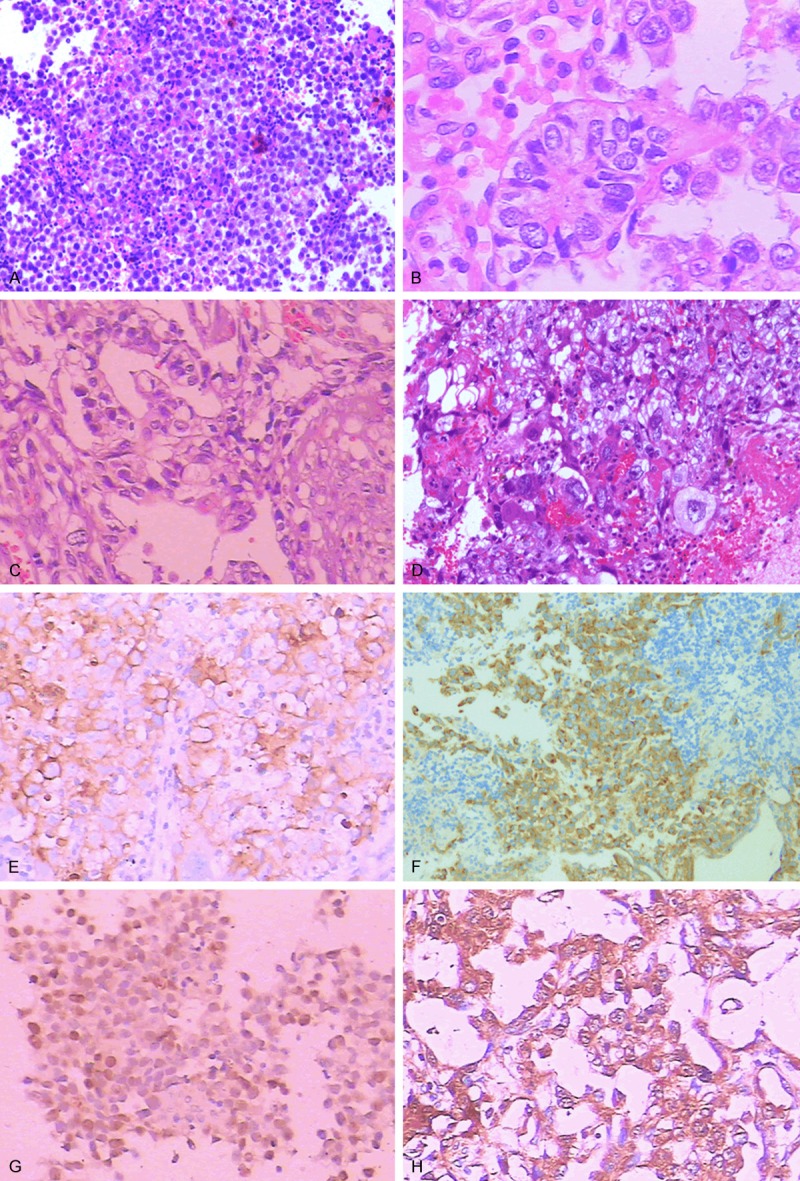
Figure 1.

Histological features: A: Germinomas (CNS) are recognized for tumor cells with abundant clear cytoplasm, sheet growth pattern, lymphocytic infiltrating along fibrovascular stroma (HE×200). B: Yolk sac tumors (mediastinum) are composed of clear, columnar epithelial cells arranged in sheets, cords and tubules structures. “Schiller-Duval bodies” is a hallmark of YST (HE×400). C: Embryonal carcinomas (CNS) are characterized by tumors cells organized in sheets, cord, or gland-like structures (HE×200). D: Choriocarcinomas (CNS) are composed of two characteristic cell types: cytotrophoblastic cells and syncytiotrophoblastic giant cells (HE×200). E: Immunostaining for PLAP in serminoma (retroperitoneum) (×200). F: Immunohistochemistry for CD117 showing positive staining in the tumor cells of the germinoma (CNS) (×200). G: Immunohistochemistry for OCT3/4 showing positive staining in the tumor cells of the germinoma (CNS) (×200). H: Immunostaining for AFP in yolk sac tumor (mediastinum) (×200).
