
Figure 3.
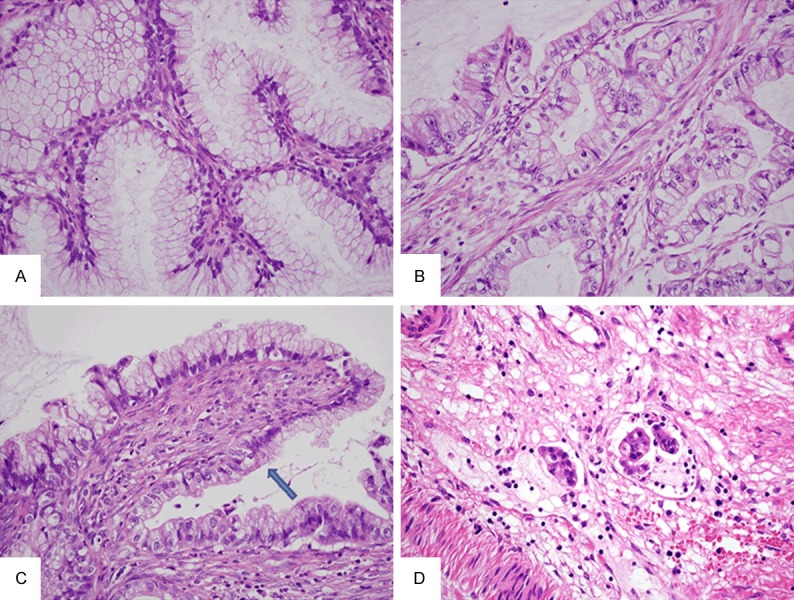
Histological findings. Irregularly shaped glands are detected. The glands are lined by single-layer columnar cells without prominent atypia. The nuclei are basally located and the cytoplasm is abundant and clear, suggesting MDA (A). Other glands contain cells with markedly enlarged atypical nuclei and prominent nucleoli. The tumor cells have abundant clear cytoplasm with prominent cell borders, reflecting the morphological features of GTA (B). Transitional area (arrow) between the MDA and GTA components is detected (C). Lymphatic involvements were observed (D).