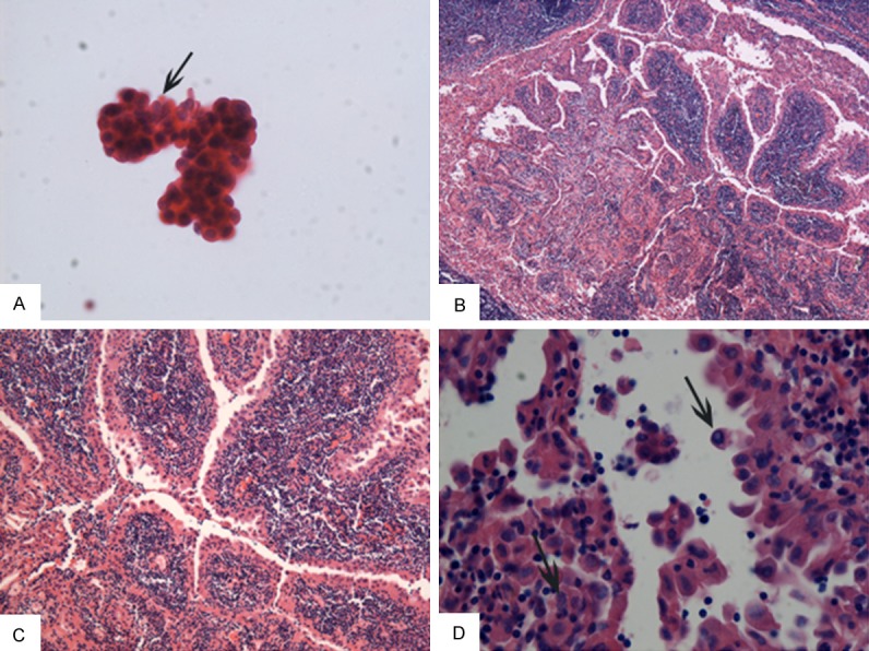
Figure 1.

Morphological examination for the case of Wathin-like papillary thyroid carcinoma (HE stain). A: Fine needle aspiration cytology. Oncocytic papillary thyroid epithelium cells with elongated nuclei, clear chromatin and nuclear grooves (arrow indicated) and scattered lymphocytes in the background (×400). B-D: Histology revealed papillae with striking lymphocytic infiltration in papillary stalks. The lining neoplastic cells exhibited eosinophilic crytoplasm and irregular, clear, overlapping nuclei with nuclear grooves (arrow) and intranuclear pseudoinclusions (arrow) (×40, ×100, ×400 respectively).
