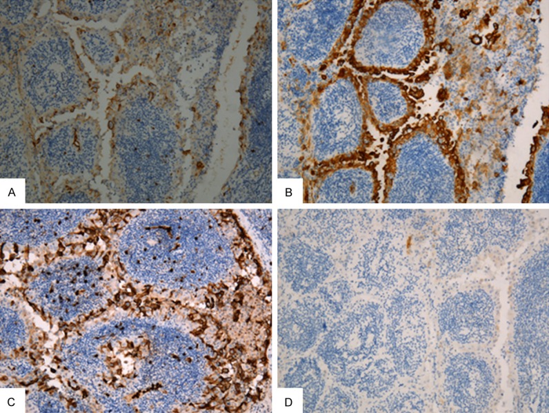
Figure 2.

Immunohistochemistry staining. Neoplastic epithelial cells show positivity at membrane or cytoplasm for HBME-1 (A), 34βE12 (B), Leu-M1 (C), respectively (×100). (D) RET/PTC expression using anti-RET rabbit serum. No signal present in the cytoplasm and membrane of neoplastic epithelial cells (×100).
