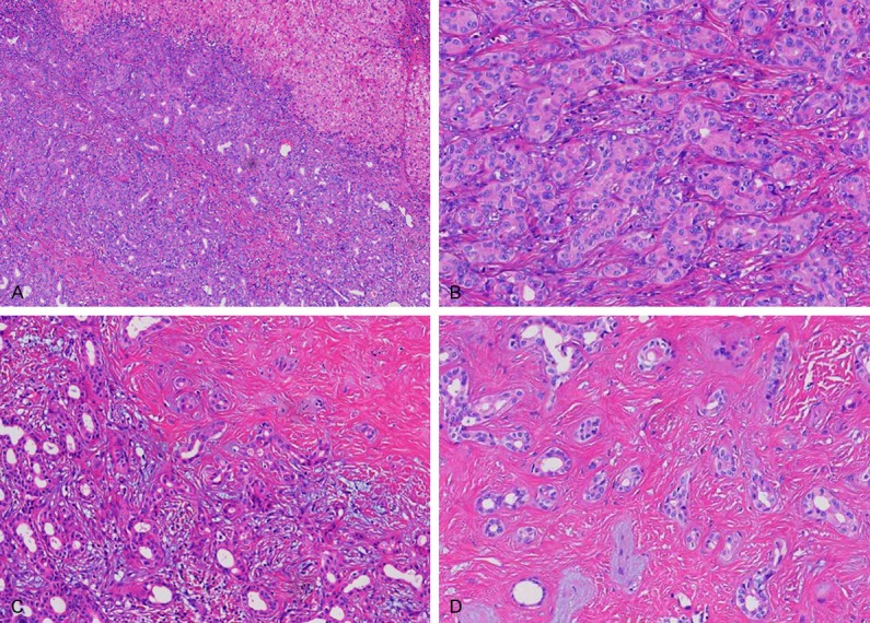
Figure 2.

Microscopically, the tumor had no capsule, but was well circumscribed from the surrounding liver parenchyma (A); the tumor was mainly composed of a proliferation of small, uniform small-sized ducts with cuboidal cells that had regular nuclei; near the periphery of the tumor was consisted of densely packed proliferation of simple tubular ducts combined with some chronic inflammatory cell infiltration (B and C); in the central fibrosis was denser, small bile duct was decreased and was separated by fibrous septa, squeezed into a slit like or irregular in shape (D).
