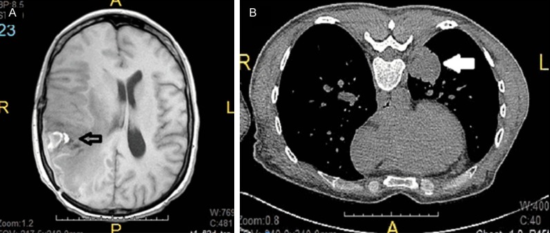
Figure 1.

Brain and lung imaging studies. A. Magnetic resonance imaging showing a 4.6 cm necrotic and heterogeneous mass at the temporoparietal junction (empty arrow). B. Computed tomography of chest showing a 3.0 cm demarcated and solid mass abutting the posterior medial pleura (white arrow).
