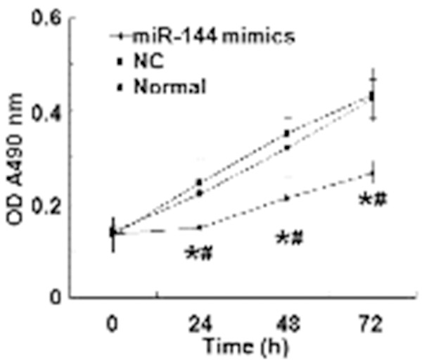
Figure 3.

Optical density of A549 cells at 24, 48, and 72 h after transfection. The proliferation of A549 cells in normal control, negative control and miR-144 mimics groups is reflected by optical density values. *, P < 0.05 compared with normal control group; #, P < 0.05 compared with negative control group.
