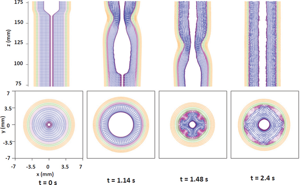
Fig. 13.
Kinematics of the esophageal layers at four different stages: at rest (t=0 s); at dilation (t=1.14 s); at contraction (t=1.48 s); at relaxation (t=2.4 s), for the Case 2 in Section 5.2. The five layers included in the model, from the inside to the outside, are the IM, OM, IF, CM, and LM layers, respectively. (Upper) Side view of a section of the esophagus within the box: (−7 mm, 7 mm) × (−0.2 mm, 0.2 mm) × (75 mm, 175 mm); (Lower) top view of a section of the esophagus within the box: (−7 mm, 7 mm) × (−7 mm, 7 mm) × (119.5 mm, 120.5 mm).

