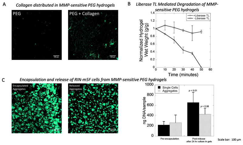
Figure 2.
A) Visualization by confocal microscopy of entrapped collagen type 1 (green) within MMP-sensitive PEG hydrogels. No collagen was detected in PEG hydrogels without collagen. B) Hydrogel degradation in the presence and absence of Liberase TL as measured by hydrogel wet weight in the absence of cells. Data are presented as the mean with standard deviation as error bars, n = 3 technical replicates) C) Rat insulinoma (RIN-m5F) cells were used as a model cell demonstrating the encapsulation of single cells and cell aggregates and their subsequent release. Encapsulated aggregates were stained with a Live/Dead membrane integrity assay (live cells fluoresce green and dead cells fluoresce red) after a 24 hour encapsulation period. Release aggregates were stained with a Live/Dead membrane integrity assay immediate after release. DNA content was used as a measure of cell number for both single cells and aggregates before encapsulation and after a 24 hour encapsulation period and subsequent release. Data are presented as the mean with standard deviation as error bars, n = 6 technical replicates). Scale bar = 100 μm. P-values are compared to pre-encapsulation.

