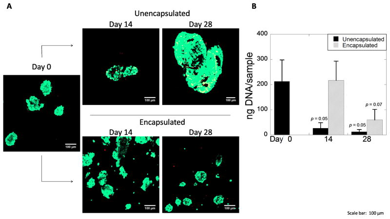
Figure 3.
A) Confocal microscopy images of aggregates of hESC-derived pancreatic precursor cells stained with the Live/Dead membrane integrity assay prior to encapsulation at day 0 (left) and after 14 and 28 days for unencapsulated (top) and encapsulated (bottom) conditions. Scale bar = 100 μm. B) DNA content as a measure for cell number at days 0, 14, and 28 for unencapsulated and encapsulated aggregates. Data are presented as the mean with standard deviation as error bars for n=3 independent experiments. P-values are compared to day 0.

