Figure 2.

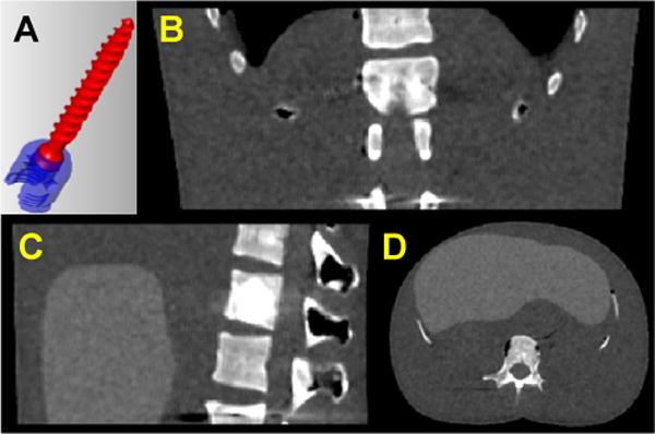
(a) CAD model of a pedicle screw. Two components of the polyaxial model are illustrated in red and blue. (b–d) Axial, sagittal, and coronal slices of the digital phantom used as a true representation of the anatomical background.

(a) CAD model of a pedicle screw. Two components of the polyaxial model are illustrated in red and blue. (b–d) Axial, sagittal, and coronal slices of the digital phantom used as a true representation of the anatomical background.