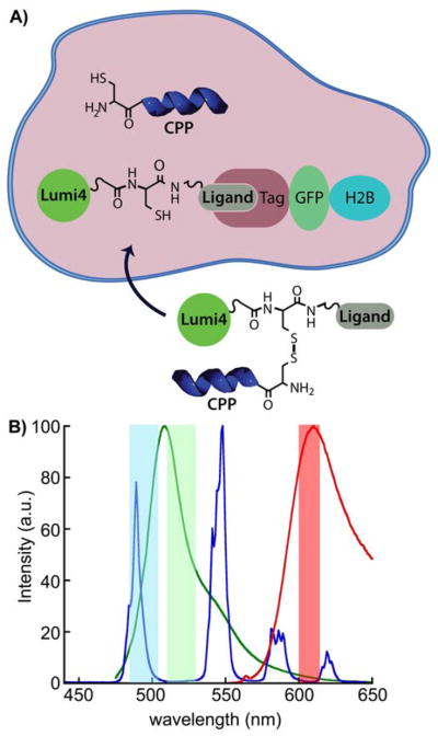
Figure 1. Model system for assessing CPP-mediated delivery and selective intracellular protein labeling.

A) Following CPP-mediated delivery into the cytoplasm, disulfide reduction frees ligand-Lumi4 heterodimer to diffuse and bind to a 3-component fusion of histone 2B, fluorescent protein and tag (eDHFR, SNAP or CLIP). Selective labeling is confirmed by observation of long-lifetime, Tb3+-to-fluorescent protein emission. B) Normalized emission spectra of TMP-Lumi4(Tb3+) (blue), EGFP (green) and mCherry (red). The characteristically narrow Tb3+ emission bands enable efficient spectral separation of donor and sensitized acceptor emission signals using narrow-pass filters (colored bands).
