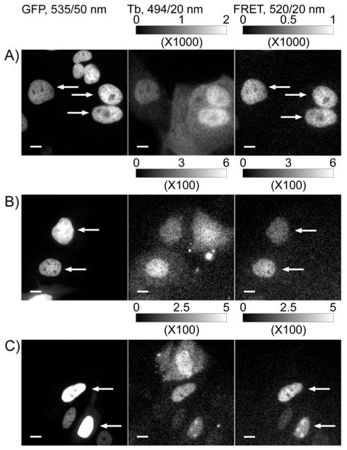
Figure 2. Conjugation to nonaarginine mediates cytoplasmic delivery of ligand-Tb3+ complex heterodimers and specific labeling of receptor fusion proteins as evidenced by time-gated imaging of Tb3+-to-GFP sensitized emission.
MDCKII cells stably expressing H2B-GFP-eDHFR or transiently expressing H2B-GFP-SNAP or H2B-GFP-CLIP were incubated with TMP-Lumi4-R9, BG-Lumi4-R9 or BC-Lumi4-R9, respectively (in DMEM w/o serum, 10 μM, 30 min), washed and imaged. Tb3+-to-GFP FRET is seen only in cells that express the target fusion protein and contain the luminescent Tb3+ complex, as indicated by arrows. A) Stable H2B-GFP-eDHFR expression; plus TMP-Lumi4-R9. B) Transient H2B-GFP-SNAP expression; plus BG-Lumi4-R9. C) Transient H2B-GFP-CLIP expression; plus BC-Lumi4-R9. Micrographs: left column, continuous wave fluorescence (λex = 480/40 nm, λem = 535/50 nm); middle column, time-gated Tb3+ luminescence (delay = 10 μs, λex = 365 nm, λem = 494/20 nm); right column, time-gated Tb3+-to-GFP FRET (delay = 10 μs, λex = 365 nm, λem = 520/20 nm). Scale bars, 10 μm. Intensity modulated displays depict the range of gray scale values in the corresponding 12-bit image.

