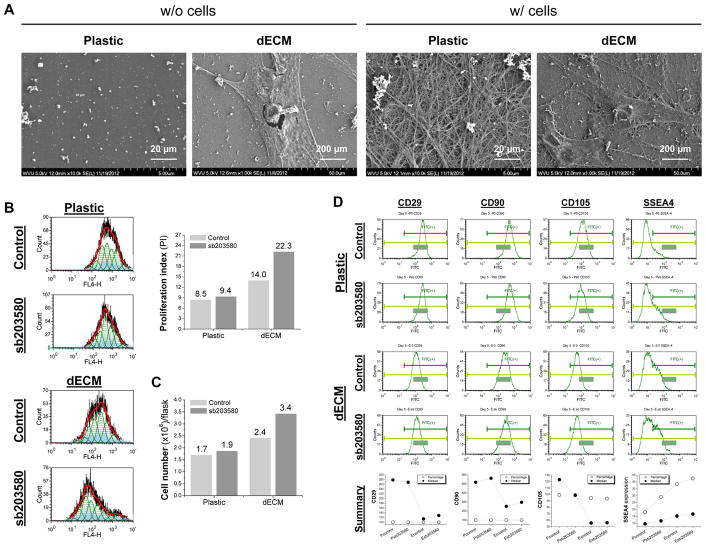
Fig. 1.
Preconditioning using sb203580 enhanced proliferation of dECM expanded hSDSCs. (A) Scanning electron microscopy was used to characterize surface topography of both Plastic and dECM (scale bar: 20 μm) and morphology of expanded hSDSCs (scale bar: 200 μm). (B) Human SDSCs were expanded on either dECM or Plastic for one passage (8 days) with or without preconditioning of sb203580. Flow cytometry was used to measure proliferation index of expanded cells. (C) A hemocytometer was used to measure cell numbers in T175 flasks (n=6) from each group. (D) Flow cytometry was used to measure both percentage and median fluorescence intensity of MSC surface markers (CD29, CD90, and CD105) and SSEA4 of expanded cells.

