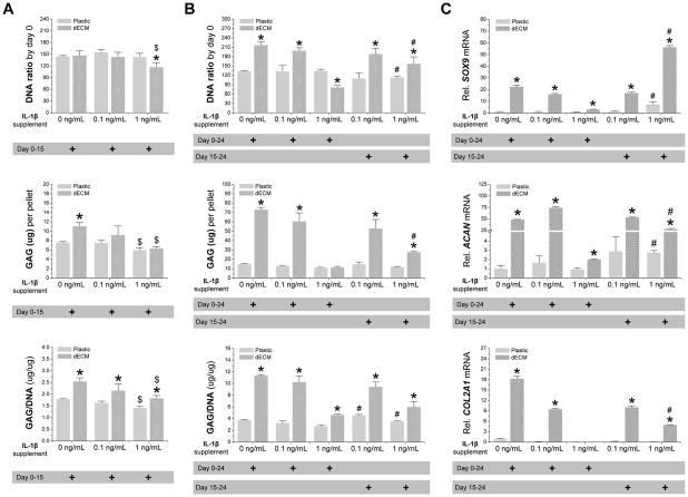
Fig. 5.
Chondrogenic induction of dECM-expanded hSDSCs in the presence of IL-1β. Human SDSCs were expanded on either Plastic or dECM for one passage followed by a pellet culture in a serum-free chondrogenic medium supplemented with either 0.1 or 1 ng/mL of IL-1β treatment for 15 days (A), 24 days, or the later stage between the 15th and 24th days (B). Biochemical analysis was used for DNA and GAG amounts in the chondrogenically-induced pellets. Cell proliferation and viability was evaluated using DNA ratio (DNA amount adjusted by that at day 0). Chondrogenic index was evaluated using a ratio of GAG to DNA. (C) Real-time PCR was used to evaluate chondrogenic marker gene expression (SOX9, ACAN, and COL2A1) in day 24 pellets. Data are shown as average ± SD for n = 4. * p < 0.05 compared with the corresponding Plastic group. # p < 0.05 compared with the corresponding group with the same dose treatment of IL-1β.

