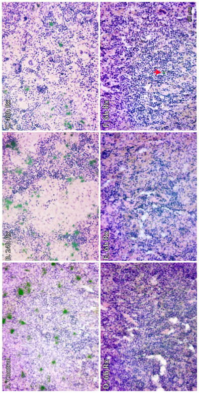
Figure 5. HIV-1 vRNA+ cells in the spleens of hu-BLT mice were detected using in situ hybridization.
Spleen tissues were collected after >70 days p.i. and fixed in 4% paraformaldehyde. Clusters of green silver grains overlay HIV-1 vRNA+ cells after radioautography for 7 day exposure. (A) Representative image showed numerous HIV-1 vRNA+ cells in the control animal (HM370). (B) Representative image showed numerous HIV-1 vRNA+ cells in Rx-24h (HM430). (C) Representative image showed numerous HIV-1 vRNA+ cells in Rx-48h (HM383). (D and E) Undetectable vRNA+ cells in two animals of Rx-6h (HM323 and 353, respectively). (F) Isolated vRNA+ cell (arrow) in Rx-6h (HM344).

