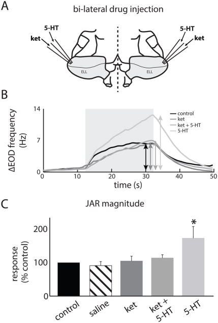
Figure 5.
A: Schematic of the experimental setup used for bilateral exogenous ket and 5-HT application in the ELL. Two electrodes containing the drugs were placed in each ELL (Materials and Methods). B: EOD frequency from an example specimen as a function of time before (black) and after drug application (ket: dark gray, ket+5-HT: medium gray, 5-HT: light gray). The double-head arrows indicate the frequency excursion while the light gray square shows the time period during which the stimulus was applied. C: JAR magnitude before application (black), after saline application (striped), after ket application (dark gray), after ket+5-HT application (medium gray), and after 5-HT application (light gray). “*” indicates that the 5-HT group is significantly different from all others at the p=0.05 level using a one-way ANOVA with Tukey post-hoc HSD (n=7). All other comparisons between group pairs were not significant (p>0.8).

