
Figure 2.
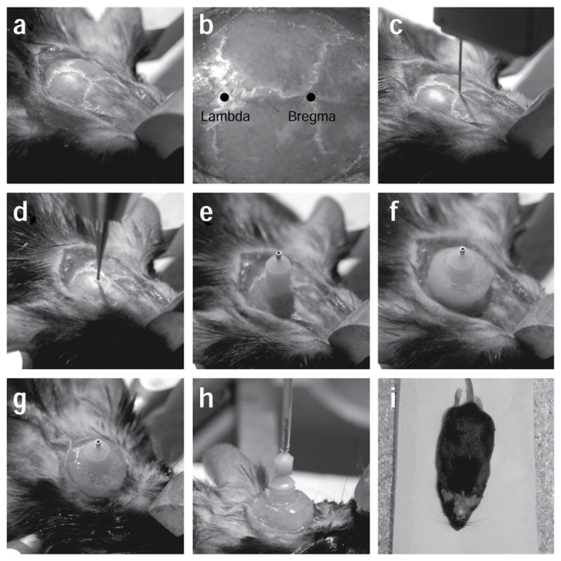
Stereotactic implantation of the cannula guide. (a) After mounting the animal into the stereotactic frame, a first incision is made to open the skin above the skull. The skin is gently pulled to the side to reveal the cranial sutures. (b) After quickly wiping the skull with hydrogen peroxide, the bregma and the lambda can be easily identified (marked spots). (c) A thin needle is used to align the skull. (d) A dental drill is used to create a small craniotomy at the desired location on the skull, without puncturing the dura. The dura is later removed using fine forceps to minimize damage to the cortex. (e) A cannula guide is implanted on the skull through the craniotomy. (f) Metabond and dental cement are used to secure the cannula guide to the skull. (g) Vetbond and sutures are used to close the incision around the cannulation site. (h) An internal cannula guide connected to a pump is inserted through the cannula guide and is used to infuse virus into the target area in the brain. (i) The animal is allowed to rest in a recovery cage after surgical implantation. The surgery was conducted according to established animal care guidelines and protocols at Stanford University.