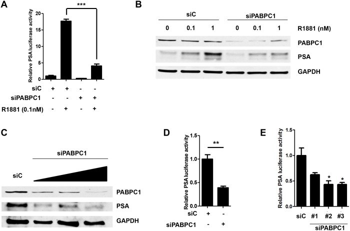
Fig 4. Knockdown of PABPC1 with siRNA decreases PSA luciferase activity and PSA protein levels.
(A) C4-2 cells were transfected with control siRNA (siC) (40 pmol/mL), siPABPC1 (pool of 3 oligos) (40 pmol/mL), pRL-CMV (0.03μg), and PSA6.1Luc (0.3μg) in OPTI-MEM. The media was changed the next day to 5% chS FBS RPMI for 24 hours followed by treatment with 0.1nM R1881for an additional 24 hours in 5% chS FBS RPMI. Luciferase assay was performed with pRL-CMV (Renilla) luciferase used as a normalizer. (B) C4-2 cells were transfected with control siRNA (siC) or siPABPC1 (40 pmol/mL) and treated as in (A) using increasing doses of R1881 (0, 0.1, and 1nM) followed by Western blot analysis. Blots were probed with antibodies specific for PABPC1 and PSA. GAPDH was used as a loading control. (C) C4-2 cells were transfected with control siRNA (siC) or siPABPC1 (pool of 3 oligos) (20 pmol/mL, 40 pmol/mL and 80 pmol/mL), in OPTI-MEM. Media was changed the next day to 10% FBS RPMI for 72 hours followed by Western blot analysis. Blots were probed as in (B). C4-2 cells were transfected with control siRNA (siC) or siPABPC1 (pool of 3 oligos) (40 pmol/mL), (D) or individual PABPC1 siRNA oligos (E), pRL-CMV (0.03μg), and PSA6.1Luc (0.3μg) in OPTI-MEM. The media was changed the next day to 10% FBS RPMI for an additional 48 hours followed by luciferase assay. Experiments were repeated three times. Significance was determined by Student’s t-test (*p<0.05, **p<0.01, ***p<0.001).

