Figure 1.

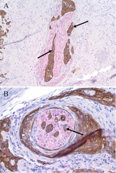
Perineural invasion in vSCC – Tumor cells (arrow) surround and invade into nearby nerves (red chromogen) in vSCC. Slides were stained using antibodies to cytokeratin AE1/3 and S100. (A) 20x magnification; (B) 40x magnification.

Perineural invasion in vSCC – Tumor cells (arrow) surround and invade into nearby nerves (red chromogen) in vSCC. Slides were stained using antibodies to cytokeratin AE1/3 and S100. (A) 20x magnification; (B) 40x magnification.