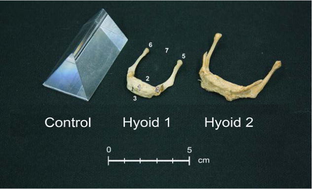
Figure 1.

Specimens scanned: Prism, Hyoid 1 (child) and Hyoid 2 (adult). Hyoid 1 is labeled to reflect anatomic landmarks listed in Table 1: (1) hyoidale, (2) hyoid body posterior superior, (3) hyoid body anterior inferior, (4) greater cornu base left, (5) greater cornu apex left, (6) greater cornu apex right, and (7) greater cornu width midpoint.
