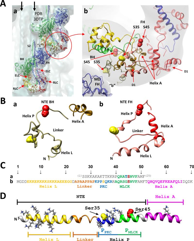
Fig. 1.
(A) Two tarantula myosin interacting-heads motifs (PDB 3DTP) are shown in (a) fitted to the tarantula thick filament 3D-map (EMD 1950, in gray).9 Both motifs interact in “b” and “c” forming part of a helix on the thick filament. The free (FH, blue) and blocked (BH, green) heads and their essential (ELC, magenta and orange) and regulatory (RLC, red/pink and yellow/tan) light chains are shown as ribbons models, and are labelled in the bottom motif. Vertical arrows: neighbour myosin tails. Bare zone: top. A zoom of the RLCs region (red circle) of (a) is shown on (b) with the blocked and free head RLCs respectively in yellow and red, with their NTEs in tan and pink. Phosphorylatable serines: Ser35 (yellow spheres), Ser45 (red spheres). Both serines of BH are located below the BH domain 1 (D1), which is located below the FH NTE9 while both FH serines are located above, freely exposed. (B) Enlarged view of the isolated blocked (a) and free (b) NTEs formed by helix L, linker and helix P, adjacent to the RLC helix A. (C) Sequence alignment of the residues 1-2529 of chicken smooth muscle RLC NTE (a) and residues 1-709 of tarantula striated muscle which included the 52-aa RLC NTE and the adjacent 14-aa helix A (b). (D) Extended model of the tarantula NTE formed by helix L (yellow), flexible coil Pro/Ala linker (orange) and helix P (blue-green), adjacent to RLC EF helix A (magenta). Helix P is formed by the target consensus sequences of PCK (PPKC, blue) and MLCK (PMLCK, green) with its phosphorylatable serines Ser35 (yellow) and Ser45 (red). Helices L and P have respectively 9 and 5 positively charged residues (white).

