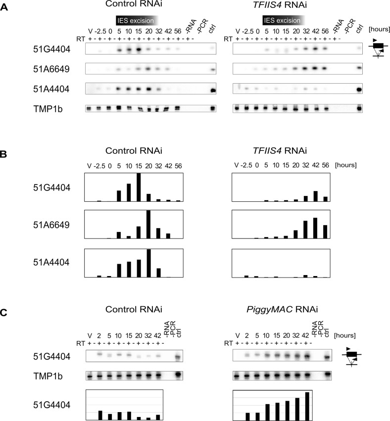
Fig 6. Detection of IES-containing (IES+) transcripts.
(A) RT-PCR and Southern blot detection of IES-containing transcripts (IES+) in a control culture (cells silenced for ICL7 gene expression) and in TFIIS4-silenced cells. Autogamy stages are marked as in S6 Fig: V–vegetative cells, -2.5 –cells during meiosis, 0 to 56 –autogamy stages in hours. Time-window when IES excision take place based on PCR shown in Fig 4 is indicated. PCR primers were located within each tested IESs: 51G4404, 51A6649 and 51A4404. The TMP1b panel shows the RT-PCR signal obtained for the constitutively expressed gene encoding trichocyst matrix protein TMP1b. (B) Histograms showing the normalization of IES+ signals shown in (A) with TMP1b mRNA. (C) Detection of IES-containing transcripts (IES+) with PCR primers located within IES 51G4404 in a control experiment, in which the ND7 gene was silenced, and in PiggyMac-silenced cells. See S8 Fig, panel B for details about autogamy stages.

