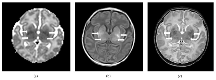
Figure 2.

Brain MRIs of a term asphyxiated newborn treated with hypothermia, performed on day 2 of life (a) and on day 9 of life (b-c). (a) The apparent diffusion coefficient map on day 2 of life for this patient shows a restricted diffusion involving the thalami and lentiform nuclei (arrows). (b and c) The T1-weighted imaging (b) and T2-weighted imaging (c) confirm the injury within the thalami and lentiform nuclei (arrows).
