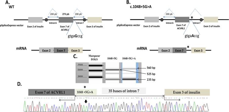
Fig 4. Splicing defect induced by the c.1048+5G>A mutation.
(A, B) Schematic construction of pSpliceExpress reporter minigene containing exon 7, Wt and mutant, and their corresponding mRNAs. The position of the c.1048+5G>A mutation is indicated with a star (on the right). The first eight nucleotides downstream of the exon-intron junction are indicated. Abnormal longer transcript generated by the new donor splice-site mutation c.1048+5G>A is shown. (C) Electrophoresis of RT-PCR products obtained after transfection in HeLa cells of normal allele, c.1048+5G (lane 2) and mutant allele, c.1048+5G>A (lane 3) revealed on TapStation (Agilent). Samples were amplified using oligonucleotides complementary to exons 2 and 3 of insulin. The uppermost band in lane 2 of 525 bp corresponds to the normally spliced wt mRNA of the exon 7 with exons 2 and 3 of insulin. The uppermost band in lane 3 of about 560 bp correponds to the aberrant transcript resulted from a splicing failure and inclusion of an intronic part in the mRNA. The lower band in lane 2 and 3 corresponds to the band containing the exons of insulin alone without the exon 7 of ACVRL1. (D) Sequencing analysis of the longer transcript resulting from the mutant construct including 35 bp of the intron 7.

