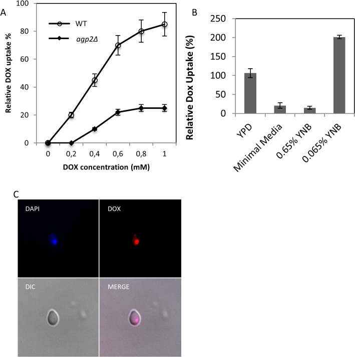
Fig 1. Relative DOX uptake into yeast cells in rich and minimal media, and localization of the drug to the nucleus.
(A)Concentration dependent uptake of DOX into the wild type (WT) strain BY4741. Cells were grown in YPD media overnight and subcultured into the same media for 1 h followed by the addition of increasing concentration of DOX and uptake was stopped after 10 min. The intracellular accumulation of DOX was monitored using FACS analysis. The agp2Δ mutant defective in DOX uptake is described below. (B)Comparison of DOX uptake in YPD, minimal media, and media containing normal amount (0.65%) of yeast nitrogen base (YNB) and 10-fold less YNB (0.065%). The WT strain was incubated in the indicated media with 800 μM of DOX for 30 min, and the intracellular accumulation of the drug was measured by FACS analysis. For panels A and B, the results were the averages of three independent analyses. (C)Epifluorescent microscopy showing intracellular colocalization of DAPI and DOX in the WT strain. The WT cells grown in YPD were transferred to low YNB followed by the uptake of DOX (100 μM) for 30 min. The fixed cells were processed for microscopy using mounting medium containing DAPI to detect the nuclear DNA. Images were captured with a DeltaVision (see material and methods). DIC, differential interference contrast; Merge, colocalization of DAPI stained nucleus with DOX.

