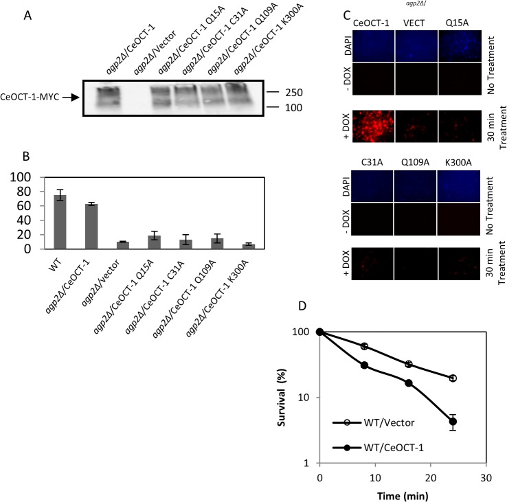
Fig 5. Expression of the native CeOCT-1, but not the variants, rescues DOX uptake in the agp2Δ mutant.
Briefly, the C. elegans oct-1 gene was cloned next to the ADH promoter and placed in frame with a C-terminal MYC epitope tag in the vector pTW438 to produce the plasmid CeOCT-1. The four variants were derived from pCeOCT-1 by site-directed mutagenesis. (A) Western blot analysis showing expression of CeOCT-1-MYC and its variants in the agp2Δ mutant. Equal amounts of total protein extracts (50 μg) were probed with anti-MYC antibodies and the molecular size markers are indicated in kD. (B) FACS analysis showing that pCeOCT-1, but not the variants, stimulates DOX uptake to nearly WT level in the agp2Δ mutant. DOX uptake was monitored using FACS analysis. (C) Epifluorescent microscopy showing that pCeOCT-1, but not the variants, causes the accumulation of DOX in the agp2Δ mutant. The cells used for this analysis were the same as for the FACS analysis in panel B. (D) Expression of CeOCT-1 sensitizes the WT cells to the killing effects of DOX. Exponentially growing cells in selective minimal media were washed twice with the low YNB uptake buffer, adjusted to OD600 of ~ 1.0 then incubated in the same buffer with 800 μM DOX in a final volume of 100 μl. Samples 20 μl were taken at 0, 8, 16, and 24 mins, diluted 10,000 fold and 100 μl plated onto solid selective minimal media to score for the surviving factions, expressed as a percentage of the zero time point set at 100%.

