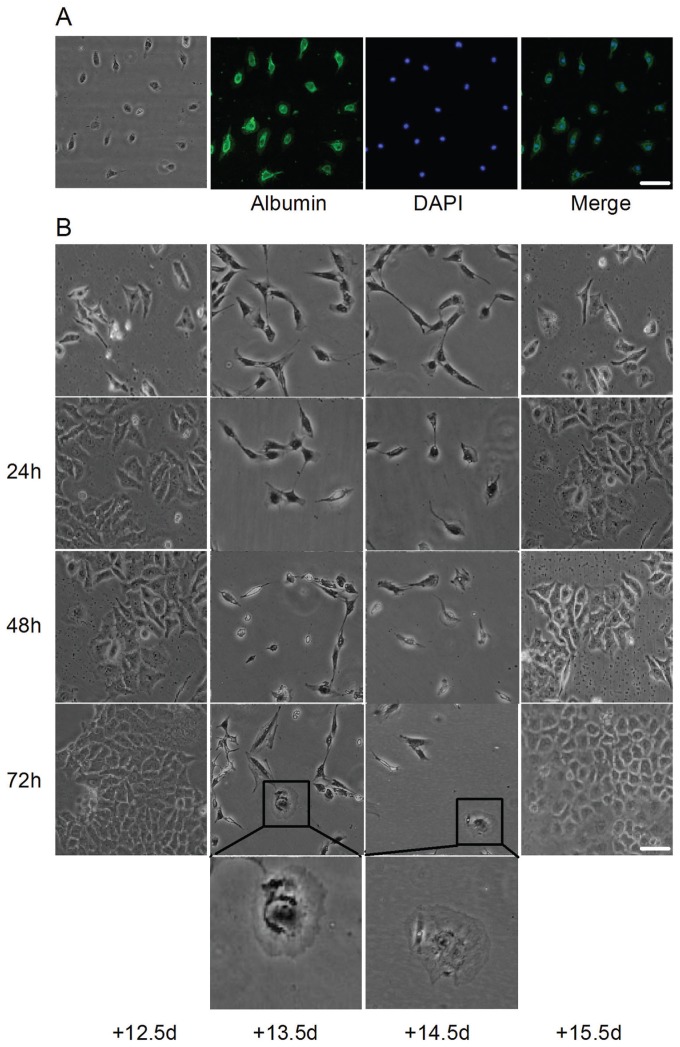
Figure 2.
Morphological changes in HepG2 cells cocultured with mouse embryonic hepatocytes. (A) Mouse primary embryonic hepatocytes were identified by detecting albumin. (B) After coculturing with mouse embryonic hepatocytes at 12.5-, 13.5-, 14.5- and 15.5-d gestation, HepG2 cells were observed. No proliferation was observed, and most HepG2 cells became round and shrunken in shape and detached and drifted in the 13.5-d and 14.5-d groups. Moreover, only a few cells were observed, and they appeared mononuclear and hexagonal, similar to hepatocytes. However, in 12.5-d and 15.5-d groups, apparent proliferation was observed with no obvious morphological changes. Scale bar = 20 μm.

