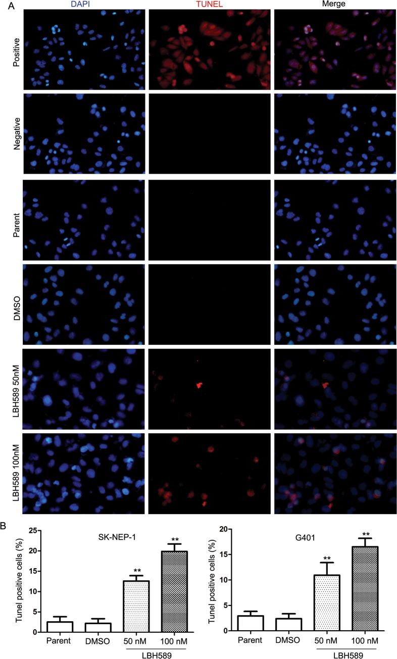
Fig 4. LBH589 induced DNA fragmentation in SK-NEP-1 and G401 cells.
The TUNEL assay relies on the presence of nicks in the DNA that can be identified by terminal deoxynucleotidyl transferase (TdT), an enzyme that catalyzes the addition of dUTPs that are secondarily labeled with a marker. Micrographs showing TUNEL staining of cells treated with LBH589 (50 nM and 100 nM) for 24 h. Red cells demonstrates the induction of DNA fragmentation. (A) The DNA fragmentation increased significantly with LBH589 treatment compared with mock treatment in both SK-NEP-1 and G401 cells. (B) Results show the percentages of TUNEL positive cells in SK-NEP-1 (100nM 19.87% ± 3.19% vs. DMSO 2.20% ± 1.99%, respectively; P = 0.003); and G401 (100nM 16.53% ± 2.86% vs. DMSO2.40 ± 1.67%, respectively; P = 0.004) cells, ** P < 0.01.

