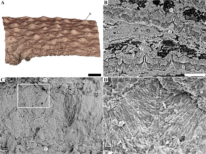
Fig 7. Eggshell morphology and microstructure of the eggs from Phu Phok.
A, 3D rendering of a portion of the surface of the eggshell of SK1-2 showing the distribution of nodes. B, tomogram of SK1-1 showing two eggshell fragments that slid in the egg, outer surfaces oriented to the top of the figure. The inner half of both shell fragments is displayed in darker shades of grey indicating the shell is less dense than the whiter outer half. Unlike micrographed thin sections (Fig 7), the funnel-shaped depression (d) do not seem to be obstructed. The pore canals (p) are highlighted by the edge interference resulting from the phase contrast effect (black and white fringes). C-D, SEM photographs of an eggshell fragment showing the fan-shaped pattern of crystal at the level of a surface node (n). Not the fibrous layer (f) underlining the eggshell. D, close up from C. Scale bars (A, B), 500 μm.

