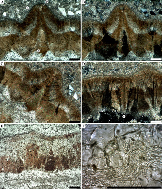
Fig 8. Micrographed radial thin section of egg SK1-5.
A-C, close up on a radial section at the level of a tall ornamentation node (n) in non-analysed polarized light (A) and analysed polarized light (B and C). The funnel shaped depression (d) exhibit similar interference colours than the innermost part of the shell. The depression tapers toward the outer surface into a very narrow pore canal (p). D, flat portion of the eggshell in analysed polarized light showing large crystals (c) with a columnar extinction. E, flat portion of the eggshell in transmitted light, showing the eggshell underlined by a fibrous layer (f). F, close up on the fibrous layer. The outer surface of the eggshell is positioned on the top part of each panels (top-right in panel C). Scale bars, 100μm.

