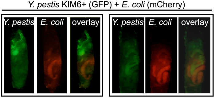
Fig 3. Localization of Y. pestis to the midgut is specific.

Larvae were coinfected with ~5x10⁸ CFU each of KIM6+ strain expressing GFP (green) and E. coli containing mCherry (red). Larvae were imaged using fluorescence microscopy to detect both GFP and mCherry and images from both fluorescent channels were overlaid. Representative images from two trials are shown (n = 15 larvae).
