
Fig. 1.
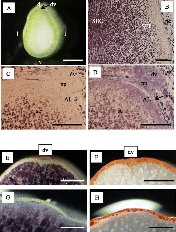
Observation of endosperm cells at 7 DAF (a–d) and in mature grain (e–h). a Median transversal section. b Microscopic observation of starchy endosperm at the center (SEC) and lateral side (SEL) stained with iodine. c, d Microscopic observation of endosperm at the dorsal side stained with iodine (c) and post-stained with toluindine blue-O (d). Matured grain stained with iodine (e, g) and Sudan IV (f, h). AL alurone cells, d dorsal side, dv dorsal vascular bundle, l lateral side, ne nucellar epidermis, np nucellar projection, pe pericarp, v ventral side. Bar: 1 mm (a), 200 μm (b–d), and 500 μm (e–h)