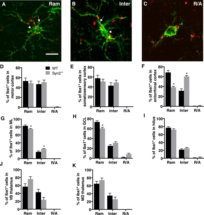
Fig 1. Microglial activation in epileptogenic 1-month old Syn2-/- mice.
Images showing Iba1/ED1 immunolabeling of microglia representing three different morphological phenotypes: ramified (Ram; A), intermediate (Inter; B) and round/amoeboid (R/A; C). Note the elongated cell soma (arrowhead) and thicker proximal processes (arrow) in B compared to A. Quantifications of the relative percentage of microglia with three different morphological phenotypes in the motor cortex (D), somatosensory cortex (E), entorhinal cortex (F), molecular layer (ML) of the dentate gyrus (G), granule cell layer (GCL) of DG (H), dentate hilus (I), ventrobasal (VB) nucleus of thalamus (J), and mediodorsal (MD) nucleus of thalamus (K) of wild type (WT) and Syn2-/- mice. Data are presented as mean ± SEM, n = 7 WT and 8 Syn2-/- mice. *, p ≤ 0.05, 2-way ANOVA with Bonferroni posthoc test. Scale bar is 10 μm (in A for A-C).

