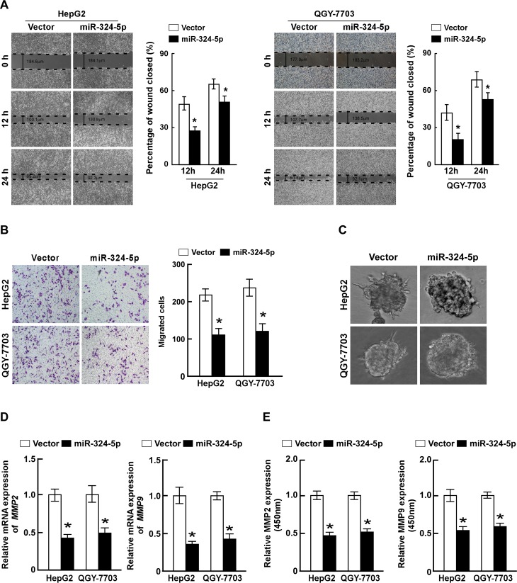
Fig 2. Ectopic expression of miR-324-5p inhibits HCC cells migration and invasion.
A. Representative micrographs (left) and quantifications (right) of wound healing assay of the indicated cells. Wound closures were photographed at 0, 12 and 24 hours after wounding. B. Representative micrographs (left) and quantifications (right) of indicated invading cells in a Matrigel-coated Transwell assay. C. Representative micrographs of indicated cells grown on Matrigel for 10 days in 3-Dimensional Cell Culture. D. Real-time PCR analysis of MMP2 and MMP9 expression in indicated cells. GAPDH served as control. E. The activity of MMP2 and MMP9 in indicated cells determined by ELISA assay. Each bar represents the mean ± SD of three independent experiments. * P <0.05.

