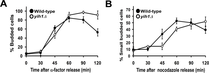
Fig 3. yih1Δ cells remain longer in the G2/M phases.

(A) Exponentially growing wild type (MSY-WT2) and yih1Δ (MSY-Y2) cells were synchronized in G1 with α-factor and released into fresh YPD media. Samples were collected at the indicated time intervals and cells were fixed. The DNA was stained with DAPI. The percentage of budded cells (mean ± S.E. of three independent experiments) was determined. (B) Cells were synchronized in metaphase with nocodazole and released into fresh YPD media. Samples were taken and stained as in A. The percentage of small-budded cells (mean ± S.E. of three independent experiments) was determined. For each time point given in the graphs, more than 300 cells were analyzed.
