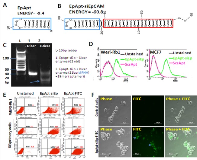
Fig 1. EpApt-siEp fabrication, in vitro processing by dicer enzyme, cell surface binding and internalization.

A. EpCAM aptamer secondary structure prediction from Mfold online. B. EpCAM aptamer siRNA chimeric construct carrying the siRNA targeting EpCAM (EpApt-siEp) is folded using Mfold online and the aptamer is indicated in blue box and the siRNA inside red box. C. EpCAM aptamer siRNA chimeric construct was incubated with the recombinant dicer enzyme at 37˚C for 18h. The reactions were performed without dicer as control reaction. Polyacrylamide gel electrophoresis of the reactions with and without dicer enzyme were run on 15% gel and stained with EtBr. The processed 21bp siRNA and unprocessed construct were observed. D. EpCAM aptamer siRNA chimeric construct was added to WERI-Rb1 and MCF7 cells in binding buffer and analyzed by flow cytometry. The overlay graph shows the uptake of the chimeric aptamer. E. Scatter plot showing the uptake of EpApt-siEp by the RB cell line, WERI-Rb1 and RB primary tumor cells. F. EpCAM aptamer siRNA chimeric construct was added to primary RB cells in media without serum for 2hr at 37˚C followed by washing with 1X PBS. Microscopic images were taken at 20X objective under phase and FITC channels of control cells alone and cells with EpApt-siEp. Data represents mean ± SD. Experiments were repeated 3 times independently with similar results. **P value of 0.01–0.001; *P value of 0.05–0.01.
