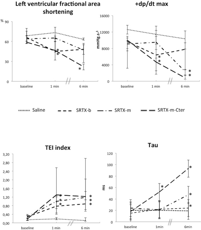
Fig 2. Effects of the toxins on left ventricular function indices.

Toxins or saline were perfused at baseline. Haemodynamic measurements were recorded at baseline, and at 1 min and 6 min post-toxin. The left ventricular area shortening and Tei index were recorded using Doppler echocardiography. +dp/dt max and Tau were recorded using a catheter placed in the left ventricle. The decrease in left ventricular area shortening and +dp/dt max indicated impairment of left ventricular systolic function. The increased Tei index reflected impairment of global left ventricular function, whereas the increase in Tau indicated impairment of left ventricular diastolic function. The dots represent the median and interquartile range. n = 15 for Saline; n = 16 for SRTX-b; n = 13 for SRTX-m; n = 12 for SRTX–m-Cter. *p<0.05 versus baseline
