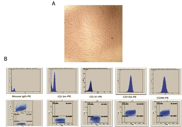
Fig.1.
Morphological appearance of cultured cells and Flow cytometric analysis. A. Mesenchymal stem cells (MSCs) showed a spindleshaped fibroblastic morphology and B. Flow cytometry analysis of the MSC surface markers. These cells were positive for CD105 and CD90 and negative for CD31 and CD34.
CD; Cluster of differentiation and PE; Phycoerythrin.

