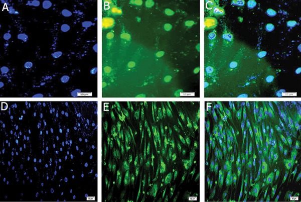
Fig.8.

A, D. Counter staining of nucleus (blue) was performed by DAPI. Expressions of of INSULIN and PDX1 in insulin producing cells (IPCs), B. FITC-conjugated PDX1 antibody detected nuclei localization of PDX1, C. Merged image of nuclei immunostaining. E. FITC-conjugated insulin antibody detected cytoplasmic localization of insulin and F. Merged (magnification ×200).
