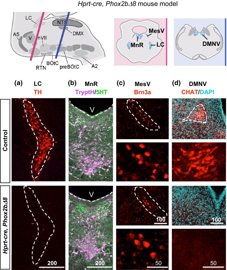
Fig. 3.
Brain pathology in Hprt-cre, Phox2b∆8 mouse. Top Cartoon of mouse hindbrain at levels of rostral (pink) and caudal (blue) hindbrain is shown. a–d The Hprt-cre, Phox2b∆8 mutant mouse showed profoundly abnormal differentiation of LC, characterized by absent expression of tyrosine hydroxylase (TH). Additional abnormalities were diminished MesV (Brn3) and DMNV Choline Acetyltransferase (CHAT) neurons. In contrast to NPARM PHOX2BΔ8 proband (see Fig. 2b), the dorsal MnR in Hprt-cre, Phox2b∆8 mouse showed non-significant reduction in counts of TrypH and 5HT cells compared with controls (n = 3). V, 4th ventricle. Scale bar unit µm

