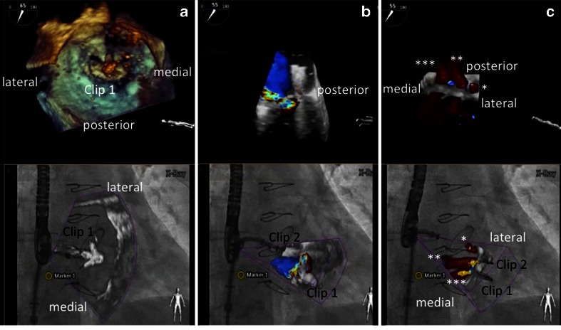
Fig. 8.
Overlay of 3D real-time echocardiography and fluoroscopy during a MitraClip procedure. a–c demonstrate step by step the insertion of clip 1 and 2 (each with 3D real-time echo on top, fusion image at the bottom). a Orientation of the clip relative to the mitral valve leaflet margin after clip opening. b Residual moderate mitral regurgitation after insertion of clip 1 in the medial part of the segments two. A second clip is approaching the valve. c Residual mild mitral regurgitation with three jets (denoted with single, double, and triple asterisks) after the insertion of the second clip

