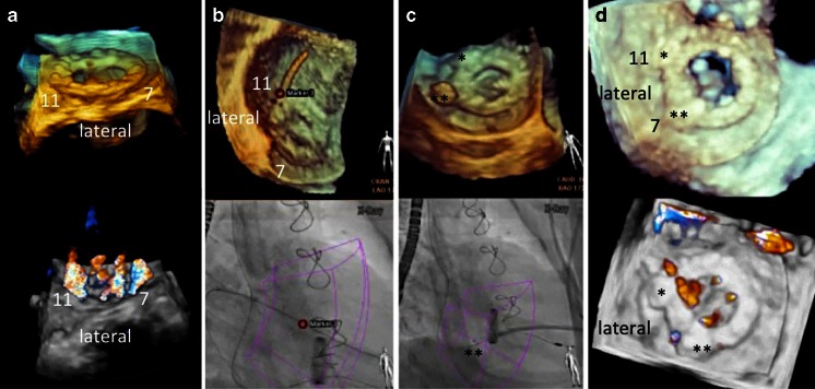
Fig. 9.
Fusion imaging during mitral paravalvular leak closure. 3D real-time echocardiography with and without color Doppler is helpful to determine the exact location and numbers of valvular and paravalvular jets (a). In this case, jets at 7 and 11 o’clock were considered the target lesions. b A red marker was used to locate the lesion at 11 o’clock (upper image), guiding the wire passage of the correct leak under fluoroscopy (bottom image). c The same approach was used to close the lesion at 7 o’clock with a vascular plug (double asterisks). d Echocardiographic images of the final result showing vascular plugs (single and double asterisks) at 11 and 7 o’clock, respectively, and a markedly reduced paravalvular and unchanged transvalvular regurgitation compared to a

