Fig. 2.
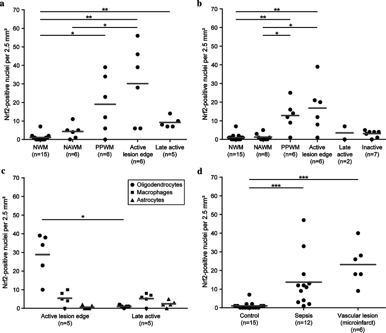
Nrf2 expression in MS lesions and controls. a, b Nrf2 expression in the normal white matter of controls (NWM), in the normal-appearing white matter of MS patients (NAWM), in the periplaque white matter (PPWM), at the active lesion edge and in the late active or inactive lesion center. Nrf2 expression is significantly increased in the PPWM and at the active edge of active demyelinating lesions in patients with acute MS and RRMS (a) and in slowly expanding lesions in patients with PPMS and SPMS (b), in comparison with normal white matter of controls. c The average numbers of Nrf2-positive oligodendrocytes, astrocytes and macrophages at the edge of active lesions and in the demyelinated (late active) lesion center are depicted. d In comparison with the white matter of unaffected control patients, Nrf2 expression is increased in control patients with concomitant microinfarcts and in control patients without pathological alteration in the brain or spinal cord, who died under septic conditions. Scatter plots depict all actual data points (black dots) as well as the median of each group (gray bars) *P < 0.05; **P < 0.01; ***P < 0.001

