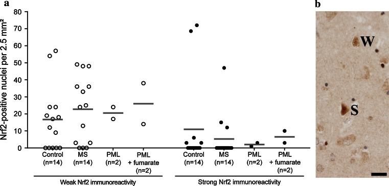
Fig. 5.
Nrf2 expression in cortical neurons. Based on the level of Nrf2 immunoreactivity (cytoplasmic and nuclear), we distinguished between two types of Nrf2-positive neurons: neurons with weak and neurons with strong Nrf2 expression. Examples are shown in (b) and labeled as weak (W) or strong (S). a Grouped according to Nrf2 immunoreactivity (weak or strong), a quantitative comparison of Nrf2-positive cortical neurons revealed no significant differences between control, MS, PML and fumarate-treated PML cases within each group. In addition to the actual data points (open and closed circles), the median of each data set is indicated by a gray bar. Scale bar 50 µm

