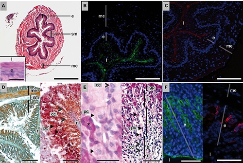
Figure 1.

Histology, histochemistry and lectin-binding of the esophagus (A–C) and stomach (D–F) of P. pipistrellus. For classical histology, HE, MT and AAS stains were used. Histochemistry was done using PAS and PAS-AB (pH 2.5) stains. Lectin histochemistry was conducted with FITC-WGA (green) and TRITC-HPA (red) and nuclei counterstain with DAPI (blue). A) HE; scale bar: 500 µm; insert: neutral carbohydrates; PAS; scale bar: 20 µm. B) Mucus in the lumen and on surface of epithelial cells bound by WGA (green) / DAPI (blue); scale bar: 200 µm. C) Mucus positive for HPA (red) / DAPI (blue); scale bar: 200 µm. D) Left: MT; scale bar: 500 µm; right: rugae; AAS; scale bar: 200 µm. E) Different cell types along the gastric gland, left: PAS; scale bar: 10 µm; right: PAS-AB; scale bar: 100 µm. F) Carbohydrate detection; left: by WGA (green) / DAPI (blue); scale bar: 200 µm; and right: by HPA (red)/DAPI (blue); scale bar: 200 µm. b, base; >cc, chief cells; e, epithelium; gp, gastric pits; i, isthmus; l, lumen; m, mucosa; me, muscularis externa; n, neck; ➤ pc, parietal cells; sm, submucosa; r, rugae; *, mucous surface cells.
