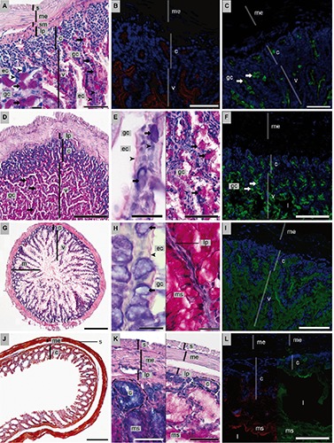
Figure 2.

Histology, histochemistry and lectin-binding of the intestine [(A–C) duodenum; (D-F) jejunum-ileum; (G-I) ileum-colon; (J-L) colon-rectum] of P. pipistrellus. For classical histology, HE and AAS stained sections are shown. Histochemistry was conducted using PAS and PAS-AB (pH 2.5) stains. Lectin histochemistry was done with FITC-WGA (green) and TRITC-HPA (red) and nuclei counterstain with DAPI (blue). A) PAS; scale bar: 100 µm; insert: PAS-positive goblet cells; scale bar: 20 µm. B) Cell surfaces positive for HPA; scale bar: 200 µm. C) WGA-positive goblet cells; scale bar: 100 µm. D) PAS; scale bar: 500 µm. E) Goblet cells positive for glycoconjugates; left: PAS-AB; mixed mucus of goblet cells, neutral glycoconjugates at luminal surfaces of enterocytes, scale bar: 20 µm and right: PAS; scale bar: 100 µm. F) Mucus and goblet cells positive for WGA; scale bar: 200 µm. G) HE; scale bar: 500 µm. H) Luminal cell surfaces of enterocytes and mucus and cytoplasm of goblet cells positive for left: PAS-AB; scale bar: 20 µm and PAS; scale bar: 40 µm. I) Goblet cells positive for WGA; scale bar: 200 µm. J) AAS; scale bar: 1 mm. K) Left: PAS-AB: crypts with acidic glycoconjugates; scale bar: 100 µm; right: PAS; scale bar: 200 µm. L) Left: epithelial cell surface of goblet cells positive for HPA and right: WGA- binding of cell surfaces and luminal mucus; scale bar: 200 µm. c, crypts; ➤ ec, enterocytes; → gc, goblet cells; l, lumen; lp, lamina propria; m, muscularis externa; ms, mucus; s, serosa; sm, submucosa; v, villi.
