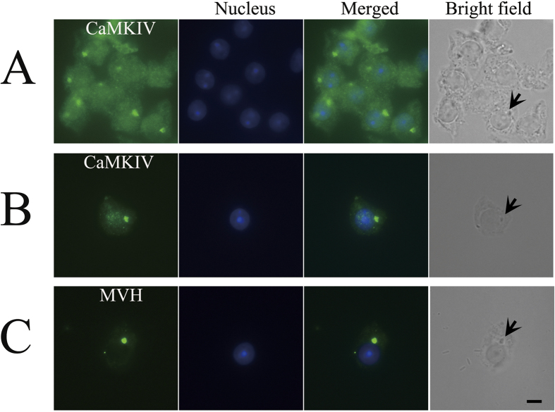
Figure 1. Localization of CaMKIV in the chromatoid body.
Testes of adult mice were subjected to squash preparation and then immunofluorescence staining. (A,B) The round spermatids were immunostained with anti-CaMKIV antibody (green). (C) Immunofluorescence staining of MVH (green). The parallel bright-field images demonstrate the location of the chromatoid bodies, which are indicated by arrows. Alexa Fluor 488 anti-rabbit IgG was used as a secondary antibody, and nuclei were stained blue with Hoechst 33342 dye. Scale bar: 5 μm.

