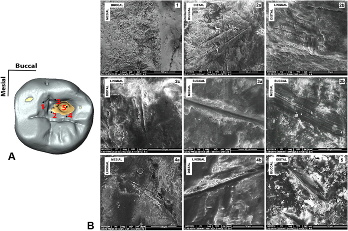
Figure 3. Scanning Electron Microscopy (SEM) images of the striations observed within the carious cavity of the Villabruna RM3.
(A) Occlusal view of the RM3 digital model, with underlined some of the areas where striations were observed. (B) The SEM images: 1, the chipping area; 2a-b-c, the mesial area; 3a-b, the buccal wall of the cavity; 4a-b, the lingual wall of the cavity; 5, inside the large carious lesion.

