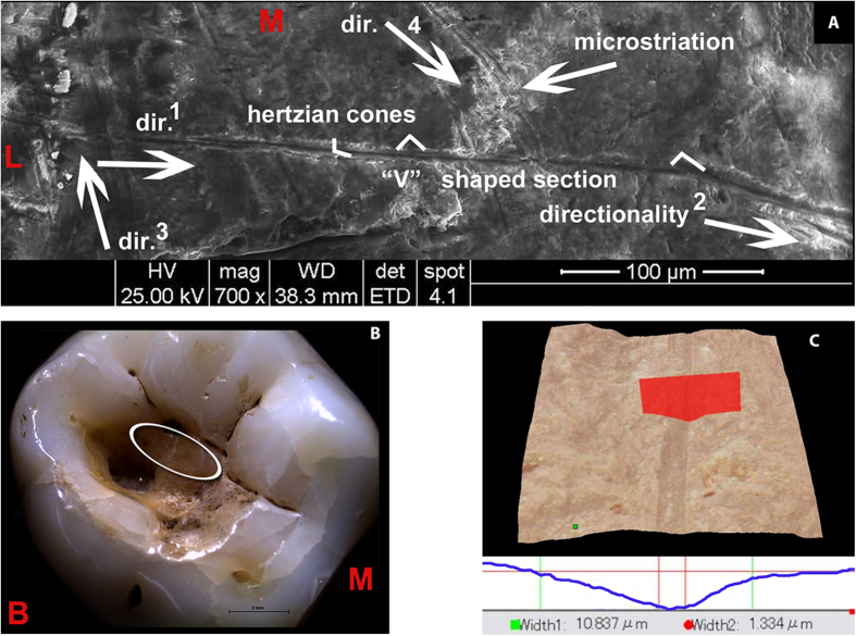
Figure 4. Morphological description of the striations observed in the Villabruna RM3.
(A) SEM images with morphological and directionality striation features (the numbers indicate the sequence of the gestures). (B) Stereo microscopical image of Villabruna RM3 with magnification of the cavity and of the region (ellipse) containing the striations described in this figure (region 2 and 4 in Fig. 3). (C) Example of 3D rendering and cross-section of the striation observed in the Villabruna tooth cavity (area 2 in Fig. 3). B, buccal; L, lingual; M, mesial.

